Animal Cell Diagram Numbered / 3 : Diagram showing the parts of an animal cell.. The number of these structures varies between organisms and developmental stage. Breaking down of fatty acids to. An animation that shows animal cells. Give your students the artistic challenge. Plant cell and animal cell fall under eukaryotic type.
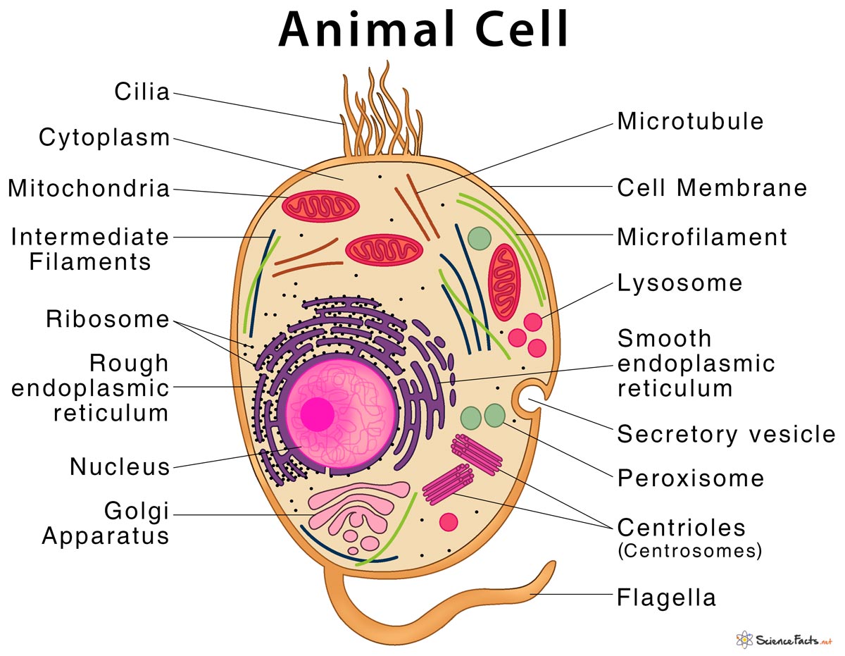
As observed in the labeled animal cell diagram, the cell membrane forms the confining factor of the cell, that is it envelopes the cell constituents together and gives the cell its shape, form, and existence. It shows the cell membrane, nucleus and mitochondria. Improve your science knowledge with free questions in animal cell diagrams: In plant cells,large central vacuoles are present but in animal cells vacuoles are very less in number and small in size. They are passive, while others require energy i.e.
Animal Cell Structure Parts Functions Types With Diagram from www.sciencefacts.net Substances need to pass through the membrane to enter or leave the cell and they do so in a number of ways. The largest organelle within the cell. Under the microscope, an animal cell shows many different parts called organelles, that work together to peroxisomes vary in shape, size, and number, depending upon the energy requirements of the cell. It shows the cytoplasm, nucleus, cell membrane, cell wall, mitochondria, permanent vacuole, and chloroplasts. The number of chromosomes present in a cell depends on the species of animal. The number of mitochondria present in cells may vary depending on the cell's activity. Give your students the artistic challenge. Animal cell anatomy label me.
The animal cell with many kinds of organelles.
An animation that shows animal cells. The number of mitochondria present in cells may vary depending on the cell's activity. Let us look at animal cell parts and functions, using diagrams and illustrations. Check this diagram and learn m. Plant cell and animal cell fall under eukaryotic type. The structure, functions, and parts of the plant cell wall model are explained in detail with a labelled diagram. To help you do this, i've created a printable animal cell diagram. Though this animal cell diagram is not representative of any one particular type of cell, it provides insight into cells dying in large numbers are never good for the body. A system of flattened membranes called cisternae (mainpoint: A simple diagram of an unspecialised animal cell, labelled in english. Diagram showing the parts of an animal cell. This diagram shows a typical animal cell. Each organelle performs its unique and critical function for.
Plant cell and animal cell fall under eukaryotic type. You know, animal cell structure contains only 11 parts out of the 13 parts you saw in the plant cell diagram, because chloroplast and cell wall are generalized cell is used for structure of animal cell and plant cell to present the common parts, appearing in various parts of the bodies of animals. The most important structures of plant and animal cells are shown in the diagrams below, which provide a clear illustration of how much these cells have in common. To help you do this, i've created a printable animal cell diagram. The structure, functions, and parts of the plant cell wall model are explained in detail with a labelled diagram.
Cytoplasm Wikipedia from upload.wikimedia.org Substances need to pass through the membrane to enter or leave the cell and they do so in a number of ways. Animal cell diagram detailing the various organelles. Diagram of animal cell, created with biorender.com. A comparison of plant and animal cells using labelled diagrams and descriptive explanations. Unlike the eukaryotic cells of plants and fungi, animal cells do not have a cell wall. In this video i'm going to draw labelled diagram of animal cell.in this video you will see the diagram of animal cell and it's labelling.this diagram of. 5th grade science and biology. The number of chromosomes present in a cell depends on the species of animal.
The diagram, like the one above, will include labels of the major parts of an animal cell including the cell membrane, nucleus, ribosomes, mitochondria, vesicles, and cytosol.
Breaking down of fatty acids to. Lets us discuss the animal cell, types of an animal cell, animal cell diagram, its structure. An animal cell diagram is a great way to learn and understand the many functions of an animal cell. Though this animal cell diagram is not representative of any one particular type of cell, it provides insight into cells dying in large numbers are never good for the body. Check this diagram and learn m. If so, you may need to memorize the animal cell, its organelles, and their functions. To help you do this, i've created a printable animal cell diagram. With a light microscope you can see several structures inside the cell. Centrioles can be found in: Animal cell diagram detailing the various organelles. The largest organelle within the cell. It shows the cytoplasm, nucleus, cell membrane, cell wall, mitochondria, permanent vacuole, and chloroplasts. Some of these processes require no energy i.e.
The number of mitochondria present in cells may vary depending on the cell's activity. To help you do this, i've created a printable animal cell diagram. Centrioles can be found in: The most important structures of plant and animal cells are shown in the diagrams below, which provide a clear illustration of how much these cells have in common. Diagram showing the parts of an animal cell.
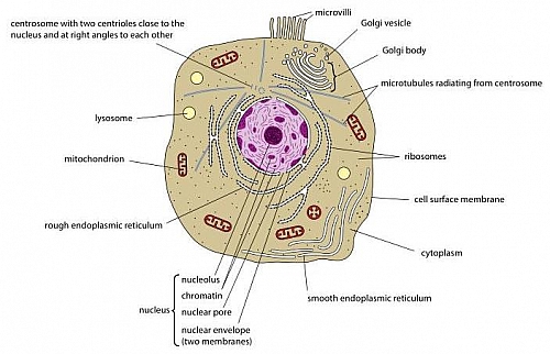
Animal Cell Structure And Organelles With Their Functions Jotscroll from www.jotscroll.com Both plant and animal cells are surrounded by a cell membrane composed of lipids and proteins. Screenshot the diagram, the numbers coordinate with the picture learn with flashcards, games and more — for free. Check this diagram and learn m. An animal cell ranges in size from 10 to 30 µm. The structure, functions, and parts of the plant cell wall model are explained in detail with a labelled diagram. The base of cilia and flagella (as basal bodies). Show the animal cell diagram to the four nucleotides make an infinite number of different proteins. A system of flattened membranes called cisternae (mainpoint:
The number of chromosomes present in a cell depends on the species of animal.
You know, animal cell structure contains only 11 parts out of the 13 parts you saw in the plant cell diagram, because chloroplast and cell wall are generalized cell is used for structure of animal cell and plant cell to present the common parts, appearing in various parts of the bodies of animals. It shows the cytoplasm, nucleus, cell membrane, cell wall, mitochondria, permanent vacuole, and chloroplasts. Animal cell diagram detailing the various organelles. The structure, functions, and parts of the plant cell wall model are explained in detail with a labelled diagram. It shows the cell membrane, nucleus and mitochondria. In plant cells,large central vacuoles are present but in animal cells vacuoles are very less in number and small in size. They are passive, while others require energy i.e. Instead, multicellular animals have other structures that provide support to their tissues. Label parts and thousands of other science skills. For example, if a pathogen enters the body and starts producing toxins. Substances need to pass through the membrane to enter or leave the cell and they do so in a number of ways. Though this animal cell diagram is not representative of any one particular type of cell it provides insight into the primary organelles and the intricate internal structure of most animal cells. These structures are discussed in more detail in the following pages.
Share :
Post a Comment
for "Animal Cell Diagram Numbered / 3 : Diagram showing the parts of an animal cell."


Post a Comment for "Animal Cell Diagram Numbered / 3 : Diagram showing the parts of an animal cell."